


For 35 years, Judith Domínguez-Cherit, MD, has worked as a dermatologist and assistant professor of dermatology at the Dr. Manuel Gea González General Hospital, the same practice her father, the distinguished dermatologist Luciano Domínguez-Soto, started in 1977.
“When I was young, my father took me with him to the hospital,” she said. “I became interested in skin diseases and was able to learn a lot of dermatology before medical school. This is why I became a dermatologist.”

Dr. Judith Domínguez-Cherit is a dermatological surgeon and an expert on the diagnosis and surgical treatment of nail diseases.
Domínguez-Cherit is a dermatological surgeon and an expert on the diagnosis and surgical treatment of nail diseases. If her significant contributions to dermatology have been obscured, this is likely due to the cultural and language barriers that can exist in medical research.
She received her medical degree from the Universidad Nacional Autónoma de México in 1982. In 1987, when she returned from Barcelona, where she had completed her residency, little was known about nail diseases. Domínguez-Cherit decided to pursue dermatological surgery because few dermatologists at the time were doing that.
“My father invited the two leading nail researchers in the world to a meeting he was hosting,” she said. This was Dr. Robert Baran, who wrote the first nail disease book in 1984 and Dr. Richard Scher, co-author of the definitive textbook on nail pathology and treatment, Scher and Daniel’s Nails (Scher and Daniel 2018). “They encouraged us to do nail studies, so I began taking biopsies.”
Since that time, she has published dozens of articles on nail diseases, including a review of the diagnosis and management of painful nail conditions (Olvera-Rodríguez et al. 2021). Domínguez-Cherit co-wrote the chapter on nail emergencies in Scher and Daniel’s Nails (Domínguez-Cherit and Gutiérrez-Mendoza 2018). She was chief of the Dermatology Department at the Instituto Nacional de Ciencias Medicas y Nutricion Salvador Zubirán (National Institute of Medical Sciences and Nutrition Salvador Zubirán) in Mexico City, and now she is working again at Hospital General “Dr Manuel Gea González”, she is a member of the National Academy of Medicine and Mexican Academy of Surgery. Domínguez-Cherit is well known throughout the dermatology community in Latin America and is the director of a nail disease course at the Latin American College of Dermatology.
While she studies numerous nail diseases, as well as other dermatological topics, her primary clinical and research interest is melanonychia.
Melanonychia and subungual melanoma
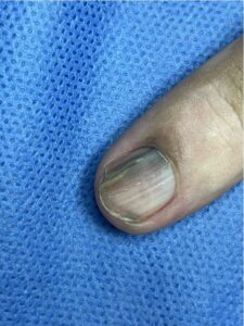
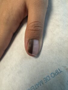
Melanonychia is a dark discoloration of the nail plate, often in a longitudinal stripe, though it can affect the entire width of a nail. It can result from trauma-induced deposits of melanin, bacterial or fungal infections, systemic diseases, medications, or subungual tumors (Domínguez-Cherit 2008). Despite the importance of determining the cause of melanonychia, there are no established treatment guidelines. Some have published diagnostic criteria (Jin et al. 2016) and developing guidelines is an ongoing focus of Domínguez-Cherit’s research (Ocampo-Garza et al. 2025). She also wrote the chapter on subungual melanomas in the textbook, Melanonychias (2017).
Subungual melanoma predominates in people from places like Africa, Latin America, and Asia. Most of the adult patients Domínguez-Cherit sees at the clinic have racial melanonychia.
The principal cause of melanonychia is activation of melanocytes — cells in the skin and eyes that produce melanin — leading to increased amounts of melanin and known as racial melanonychia. The other cause is an increase in the number of melanocytes. Nevi, lentiginous melanocytic hyperplasia, and melanoma have a larger number of melanocytes.
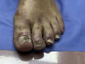
Melanonychia can be a precursor to nail unit melanoma (NUM). While generally a rare form of melanoma in white people, NUM is an aggressive cancer with poor prognosis that accounts for 20% of all melanomas in Hispanics in Mexico (Anda-Juárez et al. 2016). NUM is a subset of acral lentiginous melanoma (ALM), a cancer that presents on the palms, soles, or under the nails. ALM is rare, ranging from 1.8–2.8 per million, depending on race (Holman et al. 2023). However, it accounts for a much larger fraction of melanomas in Hispanic Black people (19.1%) compared to only 0.8% among non-Hispanic white people. ALM differs from cutaneous melanomas in that it is characterized by genomic instability, including frequent gene amplifications and chromosomal rearrangements (Yeh and Bastian 2021).
There are no effective drugs to treat NUM and, historically, treatment has been aggressive, involving digital amputation. Given that NUM predominantly affects the index finger and big toe, this can have a serious impact on quality of life.
Racial melanonychia
Melanonychia is more common, and more often leads to NUM, in those with skin of color (Ocampo-Garza et al. 2025). It occurs infrequently in those with white skin but can occur in the majority of African Americans (Singal and Bisherwal 2020). Racial melanonychia, and melanocyte activation and pigmentation, can result from the inflammation that follows nail trauma, such as friction. This is why melanonychia occurs much more often in index fingers and big toes. Continuous trauma, including friction, is necessary to diagnose racial melanonychia. Proper diagnosis and ongoing surveillance are important because melanonychia can be the first sign of melanoma in dark-skinned populations.
“When I see an adult with a longitudinal melanonychia acquired in adulthood, and that has been expanding, I assume it’s melanoma unless we demonstrate otherwise.”
“Biopsy is the definitive diagnosis,” she said. “When I see an adult with a longitudinal melanonychia acquired in adulthood, and that has been expanding, I assume it’s melanoma unless we demonstrate otherwise.”
Subungual melanoma predominates in people from places like Africa, Latin America, and Asia. Most of the adult patients Domínguez-Cherit sees at the clinic have racial melanonychia.
Saving the digit, improving quality of life
International guidelines about treatment of melanoma do not specifically differentiate NUM. Her research has shown that biopsies at the early phase of melanoma can lead to removal of the nail apparatus and preservation of the digit. Domínguez-Cherit and her colleagues published a study of 15 patients with subungual melanoma that were treated with resection of the nail unit instead of amputation (Anda-Juárez et al. 2016). Long-term follow-up (≥ 15 months) following surgery revealed no local or distant tumor recurrence, supportive of non-amputative surgery as a preferred and safe form of disease management (Ocampo-Garza et al. 2025).
The principal cause of melanonychia is activation of melanocytes, cells in the skin and eyes that produce melanin, leading to increased amounts of melanin and known as racial melanonychia.
“Our interest as dermatologists is to educate people that nail melanoma in early phases does not have to result in amputation,” Domínguez-Cherit said. “We now know that melanoma can start from a longitudinal melanonychia, can grow very slowly, and that we can prevent amputation and death from melanoma if we take care of the melanonychia.”
Even in patients with advanced melanoma involving the nail apparatus, once the tumor has become invasive, amputation of only the nail apparatus may be the preferred treatment (Nakamura Y 2014) While this does not improve outcomes, preservation of the finger or toe for the duration of a patient’s life does enhance their quality of life.
“There is no reason that people in Mexico, and all those with skin of color, need to die from acral melanoma,” she said. “It is our job to have early detection of this disease.”
Melanonychia in children is different
Domínguez-Cherit has collected nail biopsies from children to expand her research interest in nail pigmentation in that population. Racial melanonychia is not seen in children. Instead, the most common cause of pigmented nails in children are nevi (Haneke 2018). This can be confusing to an uninitiated dermatologist, especially since histology and biopsies can be suggestive of melanoma.
“We now teach students that nail pigmentation in children should not be treated as melanoma even though it looks like it,” she said. “I have seen a five mm-wide pigmented nail band grow to 10 mm in children within two years. This growth can be rapid but does not indicate melanoma. A child can have a black nail and it’s normal. But when I see a black nail in an adult, that is a melanoma.”
Continuous trauma, including friction, is necessary to diagnose racial melanonychia.
Melanonychia is important to diagnose in children so they can be followed throughout life to monitor changes in pigmentation. In adults a nail unit melanoma can take as long as 15 years to grow. First it starts as a thin longitudinal melanonichya and as the melanoma grows horizontally it spreads across the width of the nail Once this slow-growing melanoma becomes invasive, there is only a short time to cure the patient.
Her future research will study 14-15-year-olds with melanonychia, including a retrospective study of 25 children for whom she has collected nail biopsies. By following the children’s progress over a number of years, she is hoping to determine if melanoma starts in adolescence.
The role of vitamin D in dermatology
Domínguez-Cherit also studies and treats congenital nail malignment, ungual squamous cell carcinoma (SCC), and the role of vitamin D in the treatment of dermatological diseases.
She has reported that pigmented SCC can present as melanonychia (Torres et al. 2016). This cancer is rare, without treatment guidelines, and Domínguez-Cherit recommends immediate nail biopsies of new or persistent melanonychias to rule out malignancies.
She has written a review of the use of vitamin D in psoriasis, melanoma, and vitiligo, among other skin diseases (García-Galaviz et al. 2019). Her randomized, double-blind, placebo-controlled study of 65 patients with moderate-severe atopic dermatitis showed significant reduction in disease severity scores with moderate vitamin D supplementation of 5,000 IU/day (Sánchez-Armendáriz et al. 2018). This aligns with what other dermatology physician-scientists have found, such as Kurt Lu, MD and his work with high-dose vitamin D to treat sunburn and skin inflammation (Scott et al. 2017; see also the profile of Dr. Lu in the summer issue of Dermatology Focus.)
Limited funding impedes research
The Mexican government recently slashed its research budget, and the Mexican Dermatology Foundation, which offers courses, hosts congresses, and publishes a journal for its members, does not provide research funding. As a result, funding comes from the researchers themselves or from private organizations.
“Our research is done with our own money, which is why we only do clinical research,” Domínguez-Cherit said. “This makes it very difficult to do basic research, let alone molecular studies.”
“If we can publish our findings in English, our hope is that dermatologists from the US can join us, expand our studies, and make treatment guidelines for nail melanoma.”
It makes it challenging for her to get her research published in the journals most relevant to dermatologists, meaning that important clinical dermatology research is often missed by her colleagues.
“If we can publish our findings in English, our hope is that dermatologists from the US can join us, expand our studies, and make treatment guidelines for nail melanoma.”
Public and professional education
Her clinic publishes brochures to educate patients and posts on social media so people can be aware of the significance of a black line on one of their nails.
“When I meet oncologists at conferences, I try to convince them not to jump to amputation,” she said. “Making an early detection of melanoma is almost impossible for an oncologist. I’m pleased that I have been able to convince oncologists with whom I work not to amputate the digit. When I do that, I have done my work.”
References
Anda-Juárez MC, Martínez-Velasco MA, Fonte-Ávalos V, et al. Conservative surgical management of in situsubungual melanoma: long-term follow-up. An Bras Dermatol. 2016;91(6):846–848. doi: 10.1590/abd1806-4841.201645100.
Dominguez‑Cherit J, Roldan‑Marin R, Pichardo‑Velazquez P, et al. Melanonychia, melanocytic hyperplasia, and nail melanoma in a Hispanic population. J Am Acad Dermatol. 2008;59:785–791. (on file)
Domínguez-Cherit J, Gutiérrez-Mendoza D, de Anda-Juarez M, et al. Subungual Melanoma. In: Di Chiacchio N, Tosti A. (eds) Melanonychias. Springer Cham. 2017:71–84. 10.1007/978-3-319-44993-7_7.
Dominguez-Cherit J, Gutiérrez-Mendoza D. Nail Emergencies. In: Rubin AI, Jellinek NJ, Daniel CR, Scher RK. (eds)Scher and Daniel’s Nails. 4th ed. Springer Cham; 2018:383–394. doi.org/10.1007/978-3-319-65649-6_22.
García-Galaviz RA, Díaz-González JM, Cano-Aguilar LE, et al. Current use of vitamin D in dermatology. Med Cutan Iber Lat Am. 2019;47(3):170–177. doi:10.35366/91754.
Haneke E. Melanonychia. In: Scher RK, Daniel CR, eds. Scher and Daniel’s Nails: Diagnosis, Surgery, Therapy. 4th ed. Springer Cham; 2018:243–268.
Holman DM, King JB, White A, et al. Acral lentiginous melanoma incidence by sex, race, ethnicity, and stage in the United States, 2010-2019. Prev Med. 2023;175:107692. doi: 10.1016/j.ypmed.2023.107692.
Jin H, Kim JM, Kim GW, et al. Diagnostic criteria for and clinical review of melanonychia in Korean patients. J Am Acad Dermatol. 2016;74(6):1121–1127. https://pubmed.ncbi.nlm.nih.gov/26830866/
Ocampo-Garza J, Peña-Romero A, Bertolli E, et al. Non-amputative digit preservation surgery versus amputation in nail unit melanoma, a retrospective study in Mexico and Brazil. Skin Appendage Disord. 2025; https://doi.org/10.1159/000544995
Olvera-Rodríguez V, Gatica-Torres M, Carrillo-Córdova DM, et al. Painful nails: A practical approach to the diagnosis and management of painful nail conditions. Int J Dermatol. 2021;60(11):1318–1333. doi: 10.1111/ijd.15496.
Sánchez-Armendáriz K, García-Gil A, Romero CA, et al. Oral vitamin D3 5000 IU/day as an adjuvant in the treatment of atopic dermatitis: a randomized control trial. Int J Dermatol. 2018;57(12):1516–1520. doi: 10.1111/ijd.14220.
Scher RK, Daniel CR. Scher and Daniel’s Nails: Diagnosis, Surgery, Therapy. 4th ed. Springer Cham; 2018. doi.org/10.1007/978-3-319-65649-6.
Scott JF, Das LM, Ahsanuddin S, et al. Oral Vitamin D Rapidly Attenuates Inflammation from Sunburn: An Interventional Study. J Invest Dermatol. 2017;(10):2078–2086. doi: 10.1016/j.jid.2017.04.040.
Singal A, Bisherwal K. Melanonychia: Etiology, Diagnosis, and Treatment. Indian Dermatol Online J. 2020;11(1):1–11. https://journals.lww.com/idoj/fulltext/2020/11010/melanonychia__etiology,_diagnosis,_and_treatment.1.aspx(On file)
Gatica-Torres M, Arguello-Guerra L, Manuel Ruiz-Matta J, Dominguez-Cherit J. Subungual pigmented squamous cell carcinoma presenting as a grey longitudinal melanonychia in a young patient. BMJ Case Rep. 2016;2016:bcr2016215390. doi: 10.1136/bcr-2016-215390. (On file)
Yeh I, Bastian BC. Melanoma pathology: new approaches and classification. Br J Dermatol. 2021;185(2):282–293. doi: 10.1111/bjd.20427. (on file)