Challenging Cases That Have Taught Me Oncology
Jennifer Choi, MD
Professor of Dermatology Chief, Divisions of Oncodermatology and Medical Dermatology, Northwestern University Feinberg School of Medicine, Robert H. Lurie Comprehensive Cancer Center
June 2024

Dr. Choi presented three oncodermatology cases that demonstrate clinical pearls for the diagnosis and treatment of oral graft versus host disease (GVHD), nutritional deficiencies, and aleukemic leukemia cutis (LC).
Case #1
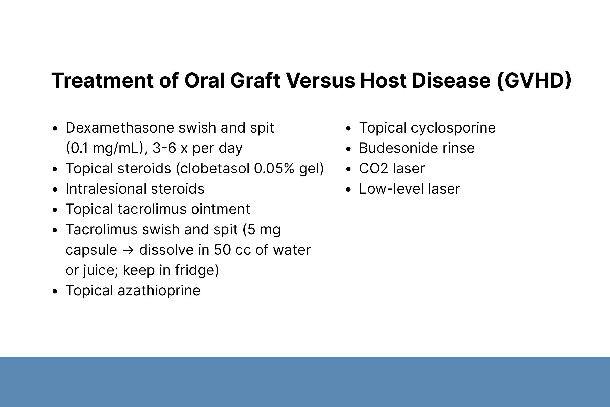
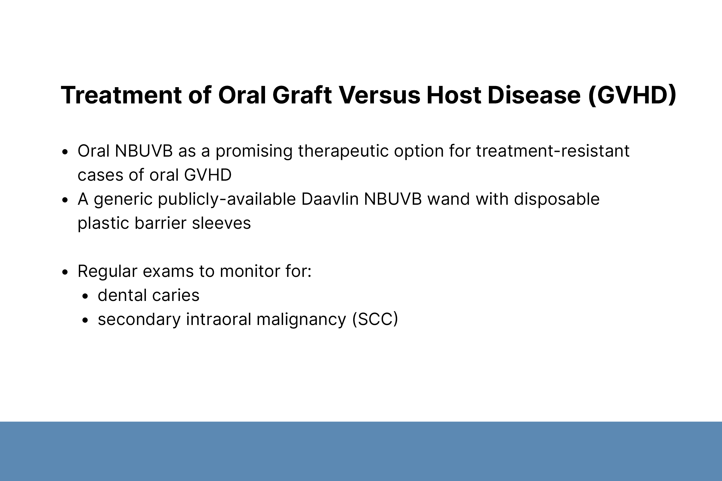
A 71-year-old female with acute lymphoblastic leukemia and hematopoietic stem cell transplant (HSCT) presented with oral pain and bleeding and bruising of the gums and oral mucosa, leading to difficulty eating and weight loss. She was diagnosed with oral GVHD but was unresponsive to first-line treatments. Because phototherapy is used to treat cutaneous GVHD, Dr. Choi treated the patient with intraoral narrowband UVB phototherapy. This resulted in complete re-epithelialization of oral erosions and significant subjective improvement.
Dr. Choi discussed the etiology, clinical presentation, diagnosis, and treatment of oral GVHD, seen in 45–90% of chronic GVHD cases. It can lead to decreased oral intake, increased oral infections, and decreased quality of life. First-line treatments include dexamethasone rinse, topical and intralesional steroids, topical tacrolimus, and compounded tacrolimus rinse. Dr. Choi presented evidence demonstrating the effectiveness of intraoral phototherapy for oral GVHD.
Case #2
A 60-year-old female with metastatic lung adenocarcinoma and an epidermal growth factor receptor (EGFR) mutation was treated with the EGFR inhibitor osimertinib for two years. After disease progression, she was treated with adjuvant intravenous amivantamab, a bispecific antibody against EGFR and mesenchymal-epithelial transition (MET) receptor.
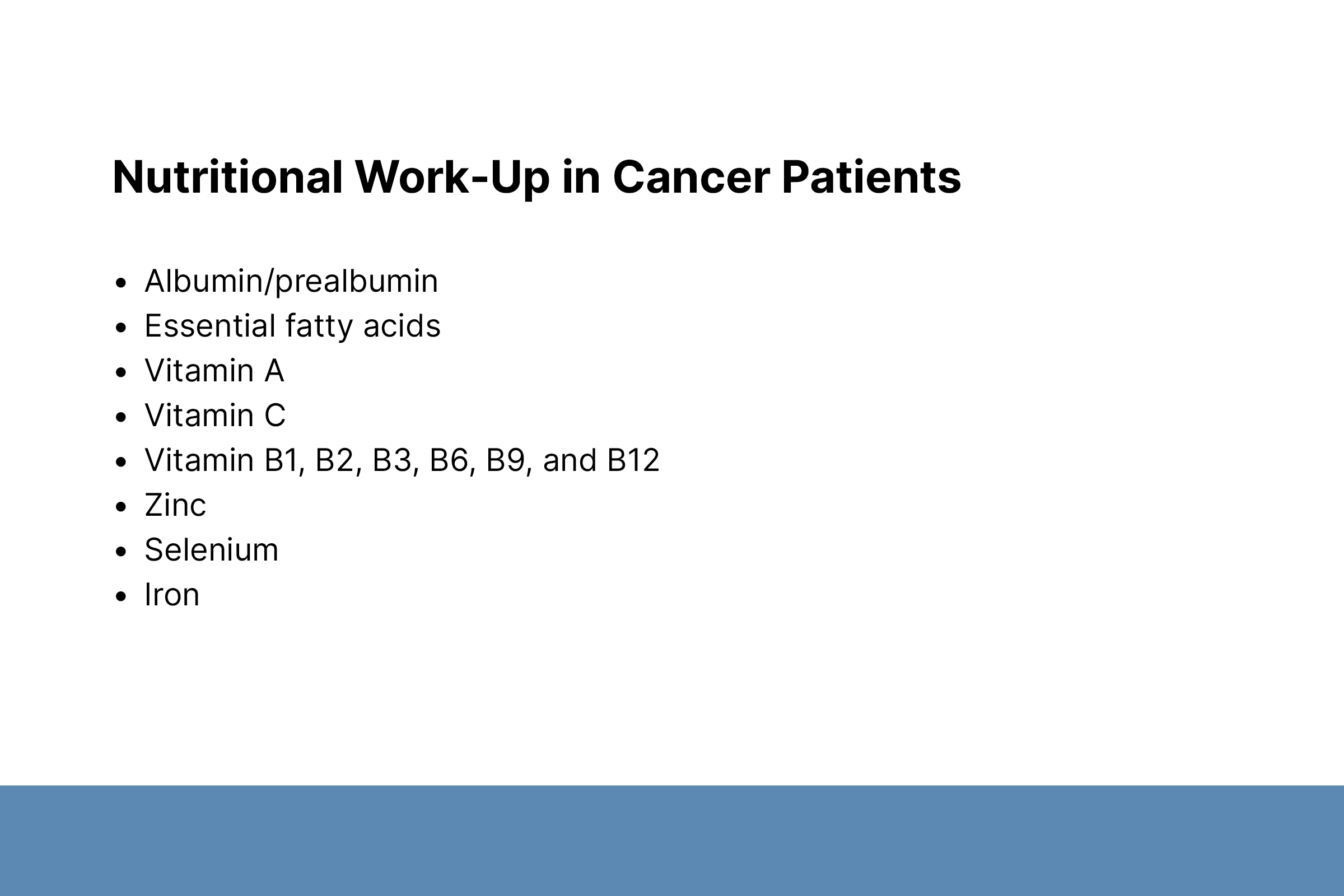
The patient presented with dry, itchy skin after two amivantamab infusions. She was unresponsive to multiple skin-directed treatments. Tissue culture identified Pseudomonas aeruginosa infection. Treatment with oral antibiotics was helpful but not completely sufficient. Dr. Choi checked the patient’s nutritional status and identified prealbumin and zinc deficiencies. The patient healed quickly soon after by starting a zinc supplement and eating a high-protein diet.
Dr. Choi described cutaneous reactions to EGFR inhibitors, which occur in 45–90% of patients. Superinfections occur in 30% of cases and require tissue culture to diagnose.
Dr. Choi noted the importance of continuing to search for a reason when a patient is unresponsive to treatment. She encouraged dermatologists to consider nutritional status in skin healing because cancer patients have a higher risk for nutritional deficiency. She credits her colleague Dr. Bernice Kwong at Stanford for first highlighting this need in oncology patients.
Case #3
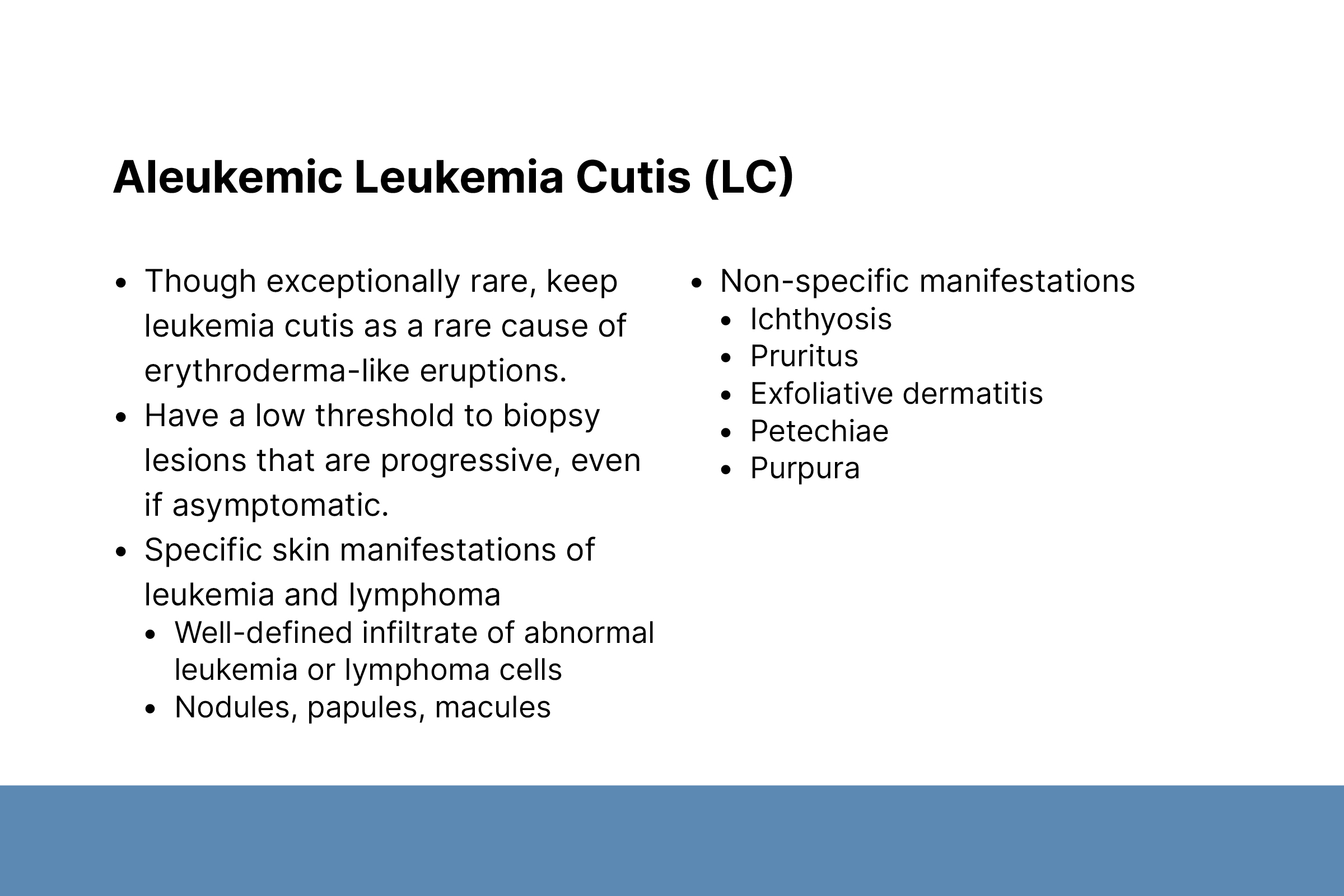
A 50-year-old female was treated for acute myeloid leukemia and subsequently developed a diffuse eruption. Biopsy result was consistent with widespread aleukemic leukemia cutis mimicking erythroderma. The eruption completely cleared after treatment with high-dose cytarabine.
Dr. Choi discussed the etiology, clinical presentation, and diagnosis of aleukemic LC, which occurs secondary to leukemia. Aleukemia LC occurs in 5–7% of LC cases when malignant cells invade the skin before the blood or bone marrow. The condition has a poor prognosis. Dr. Choi suggested a low threshold to biopsy asymptomatic, progressive lesions and emphasized that aleukemic LC should be treated like leukemia.